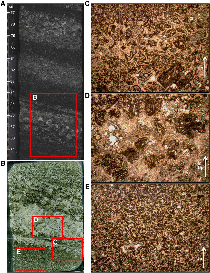
Figure F17. (A) Core section image, (B) thin section scan, and photomicrographs (plane-polarized transmitted light) of sparsely vesicular vitric tuff in Section 324-U1348A-18R-1, top of Unit III, with (C) glass shards and bubble wall fragments with elongate vesicular shard at bottom, (D) subrounded vesicular basalt clasts, and (E) fine hyaloclastite matrix. Thin section scan location of B is indicated by the red box in A. Width of field of view of photomicrographs is ~15 mm (1.25x) in panels C, D, and E. Yellow arrows = orientation in core.
Previous | Close | Next | Top of page