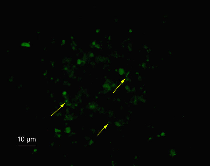
Figure F54. Microscopic image of acridine orange–stained sediment slurry (45 mbsf) after ultrasonication and sedimentation of particulate matter. Microbial cells (yellow arrows) can be distinguished from other particles.
Previous | Close | Next | Top of page