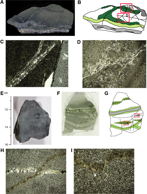
Figure F307. Vein morphologies. A. Shear vein developed in an altered chilled margin (Thin Section 33; Sample 312-1256D-194R-1, 8–13 cm) (cross-polarized light). B. Sketch of A indicating areas of C and D. C. Splayed vein in A (field of view [FOV] = 2.25 mm). D. Crosscutting and diffuse veins in A (FOV = 4.5 mm). E. Vein network with a wide vein (interval 312-1256D-205R-1, 11–14 cm). F. Whole-thin section scan of E. G. Sketch of E, with areas of H and I indicated. H. Quartz vein cut by amphibole vein (Thin Section 49; Sample 312-1256D-205R-1, 10–14 cm) (cross-polarized light; FOV = 4.5 mm). I. Amphibole vein cut by a thin vein with similar composition (Thin Section 49; Sample 312-1256D-205R-1, 10–14 cm) (cross-polarized light; FOV = 2.25 mm).
Previous | Close | Next | Top of page