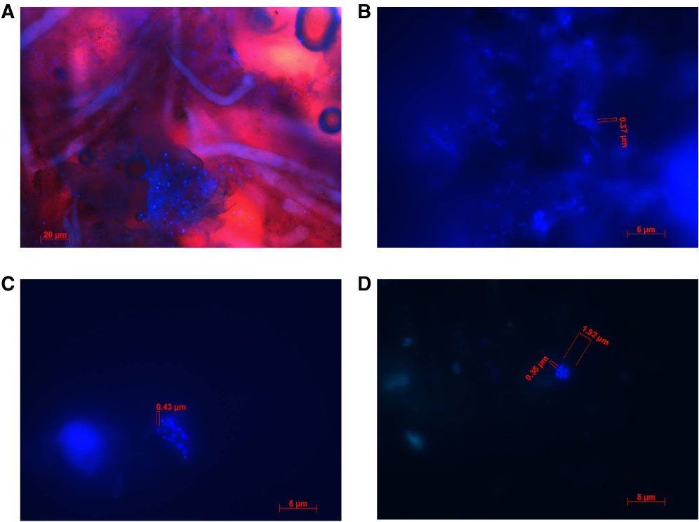
Figure F3. DAPI-stained samples under fluorescence microscopy. A. Eukaryotic organisms (e.g., sponges) and embedded clusters of microbes (blue dots) (Hole M0020A, 0.41 mbsf). B. building microbial associations (blue dots) (Hole M0020A, 4.51 mbsf). Individual cells are small (diameter = ~0.4 µm). C. Biofilm building microbial associations (blue dots) (Hole M0020A, 15.49 mbsf). Individual cells are small (diameter = ~0.4 µm). D. Seven-cell aggregates of coccoid microbes (blue dots) (Hole M0023A, 0.12 mbsf). Individual cells are small (diameter = ~0.35 µm). Cells were abundant in this sample. (Continued on next two pages.)
Previous | Close | Next | Top of page