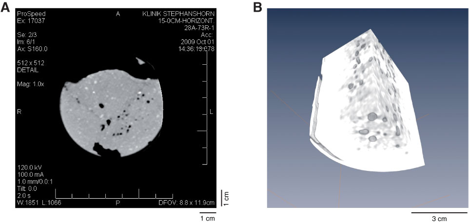
Figure F7. A. X-ray CT image of a 1 mm transverse slice in sedimentary core made during Expedition 313. Different mineral components in sand can be identified, and quantitative estimation of total sediment density is also possible. B. 3-D image block (AMIRA software).
Previous | Close | Next | Top of page