Previous | Close | Next
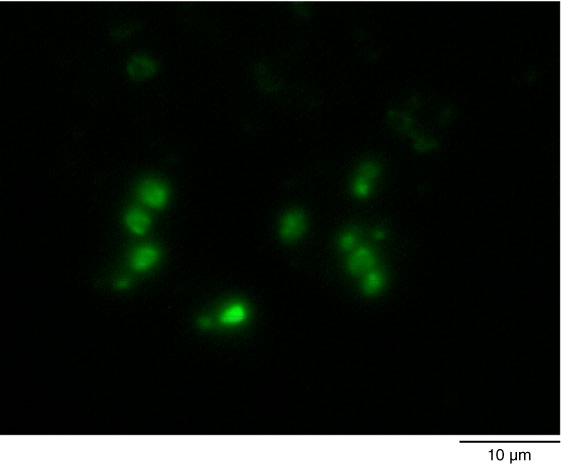
Figure F50. Fluorescent microscopic image of SYBR Green I–stained cells detected from the gravel layer (Sample 316-C0007D-17H-1, 157.7 m CSF).
Previous | Close | Next | Top of page