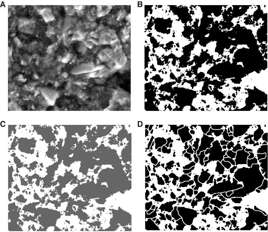
Figure F6. Illustrations of steps used during image analysis of microfabric (Sample 316-C0006E-5H-1, 128 cm). A. Environmental scanning electron microscope image. B. Binary image obtained with ImageJ software. C. Binary image (transparency = 60%) overlying the original image using CorelDraw software. D. Binary image after particle separation using the eraser tool in CorelDraw.
Previous | Close | Next | Top of page