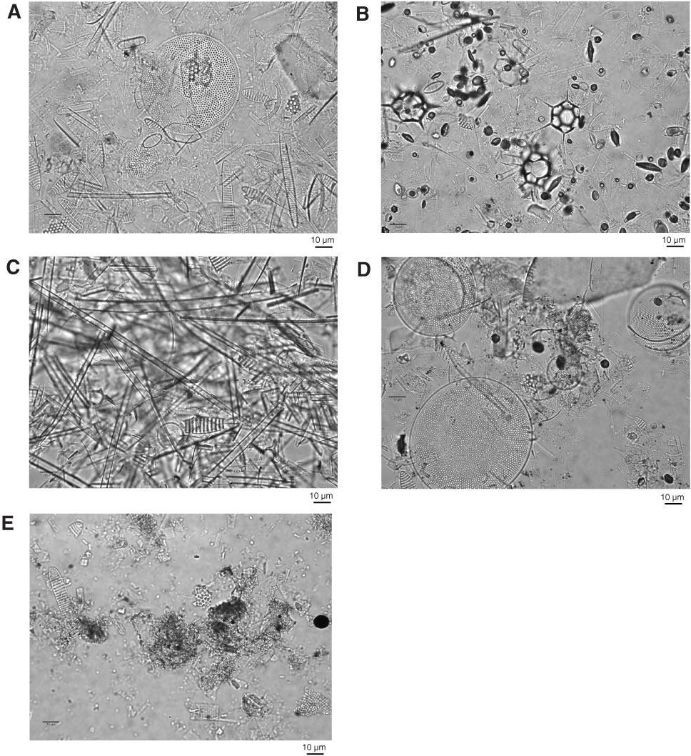
Figure F18. Photomicrographs of representative fields of view from millimeter- to centimeter-scale laminations, Hole U1357A. A–C. Biosiliceous-rich laminations; (A) gray fibrous lamina with large centric diatom Thalassiosira lentiginosa, a number of Fragilariopsis (pennate) species, and Chaetoceros resting spores (Sample 318-U1357A-19H-4, 147 cm; 147.00 mbsf); (B) orange lamina with multiple specimens of the silicoflagellate Distephanus speculum speculum (hexagonal structures) and numerous Chaetoceros resting spores (small dark shapes) (Sample 318-U1357A-19H-1, 86 cm; 169.96 mbsf); (C) white lamina with needle-shaped pennate diatoms Thalassiothrix antarctica (narrower pennate) and Trichotoxon reinboldii (wider pennate) (Sample 318-U1357A-19H-2, 19 cm; 170.78 mbsf). D. Olive-green siliciclastic lamina with several species of Thalassiosira (centric diatoms), fragmented pennate diatoms, and Chaetoceros resting spores (Sample 318-U1357A-19H-2, 80 cm; 171.39 mbsf). E. Black siliciclastic lamina with siliciclastic components and several fragmented pennates (Sample 318-U1357A-19H-6, 45 cm; 177.04 mbsf).
Previous | Close | Next | Top of page