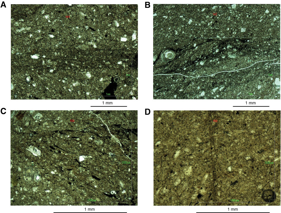
Figure F27. Microstructure of cuttings samples. A. Vein structure in plane-polarized light, Sample 319-C0009A-141-SMW (1352.7 m MSF). B. Vein structure highlighted by opaque minerals (predominantly pyrite), Sample 319-C0009A-142-SMW (1357.7 m MSF). C. Vein structure with preferred orientation of opaque minerals, Sample 319-C0009A-156-SMW (1422.7 m MSF). D. Tip of thin vein structure with local fracture, Sample 319-C0009A-156-SMW (1422.7 m MSF).
Previous | Close | Next | Top of page