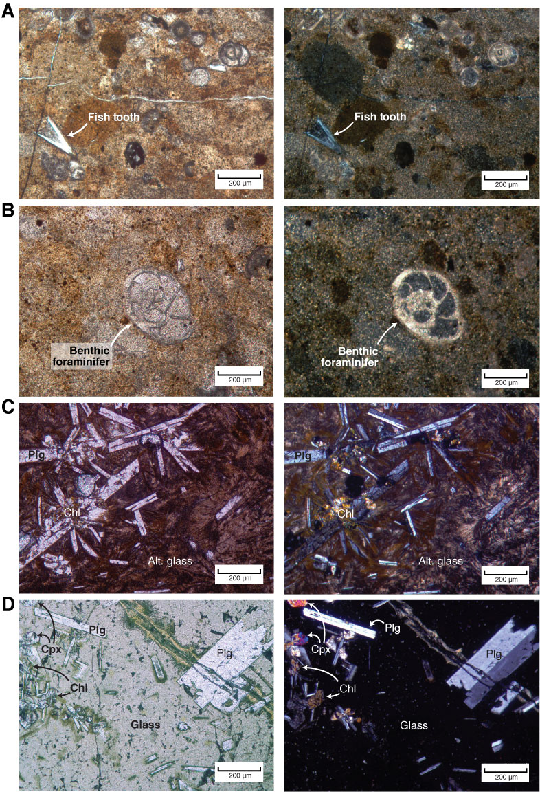
Figure F7. Thin section photomicrographs. Left image = plane-polarized light, right image = cross-polarized light. A, B. Conglomerate (Sample 320-U1331B-10H-6, 120–128 cm). Foraminifers, fish teeth, and coarse mud clasts (up to 2 cm diameter) are visible. Calcareous benthic foraminifer with calcite test walls but chambers in-filled by diagenetic silica shown in B. C. Interior of basement basalt (Sample 320-U1331A-22X-CC, 37–44 cm). D. Chilled margin of basement basalt, same sample as C. Glass matrixes are comparatively fresh. Plg = plagioclase feldspar, Cpx = clinopyroxene, Chl = chlorite, Alt. glass = altered glass.

Previous | Close | Next | Top of page