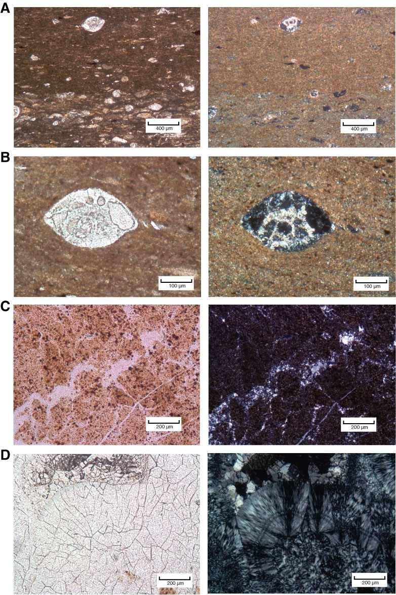
Figure F6. Thin section photomicrographs of porcellanites, Site U1332. Left image = plane-polarized light, right image = cross-polarized light. A, B. Porcellanite with foraminifers and coarse basal layers (Sample 320-U1332A-15X-2, 112–114 cm). C, D. Porcellanite with veins of recrystalized silica (Sample 320-U1332A-17X-1, 0–4 cm). Large radial crystals of chalcedony are visible in D.
Previous | Close | Next | Top of page