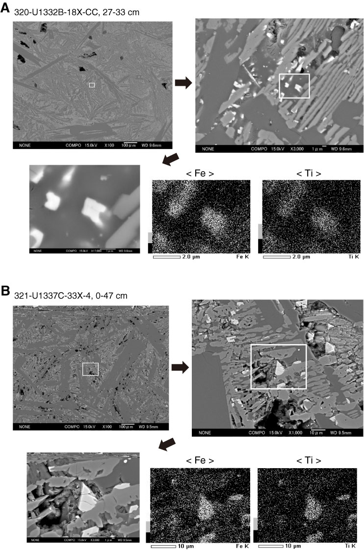
Figure F5. Backscattered electron microscope images of Samples (A) 320-U1332B-18X-CC, 27–33 cm, and (B) 321-U1337C-33X-4, 0–47 cm. Three different magnifications are shown for each. Fe and Ti elemental maps obtained by energy-dispersive X-ray spectroscopy are also shown for the highest magnification observations.
Previous | Close | Next | Top of page