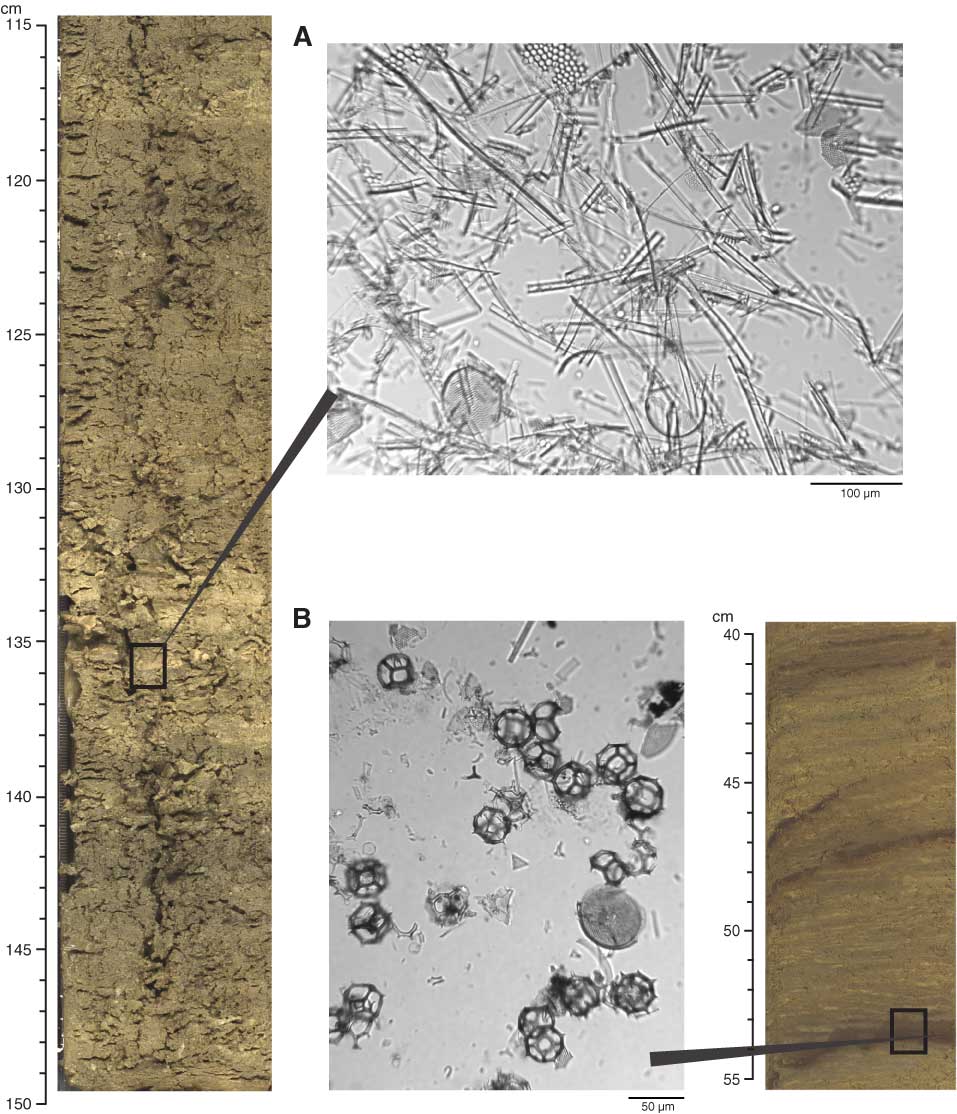
Figure F10. Core photographs and photomicrographs of smear slides of laminations. A. Nearly monospecific diatom ooze lamina with Lioloma pacificum from interval 323-U1340A-52H-5, 115–150 cm. B. Silicoflagellate-rich lamina from interval 323-U1340A-6H-7, 40–55 cm. Boxes indicate the approximate position of smear slide sample locations.
Previous | Close | Next | Top of page