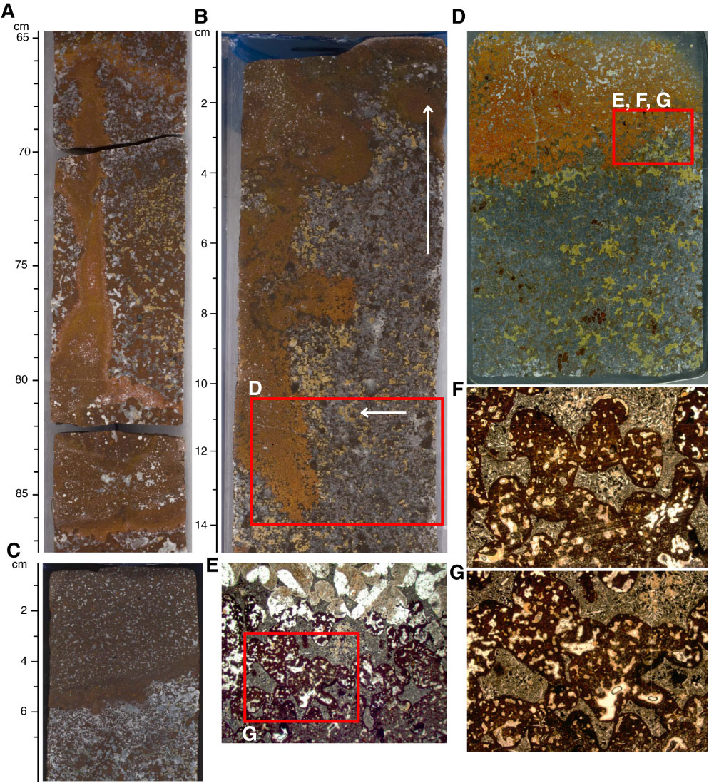
Figure F20. Core section images of Sections (A) 324-U1349-11R-3, (B) 11R-5, (C) 11R-4, (D) thin section scan, and (E–G) photomicrographs (plane-polarized transmitted light) of lava mixing features in Core 324-U1349A-11R, top of Unit IV. Thin section scan location of D is indicated by the red box in B. Red box in D indicates photomicrograph location. G is a close-up view of the area indicated by the red box in E. Field of view for D is 36 cm x 54 cm. Width of field of view of F and G is ~6 mm (2.5x) and E is 15 mm (1.25x).
Previous | Close | Next | Top of page