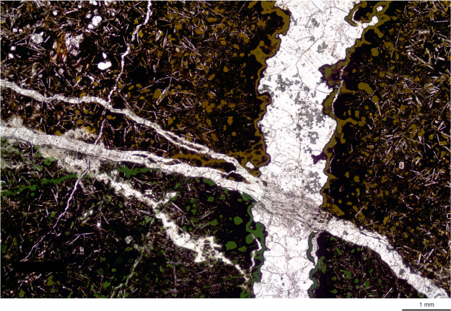
Figure F29. Photomicrograph of crosscutting calcite veins with boundary between gray and green alteration visible. The near-diagonal calcite vein has acted as a barrier for the alteration fluids responsible for brown alteration (Thin Section 51; Sample 324-U1346A-14R-2, 66–70 cm). Transmitted light.

Previous | Close | Next | Top of page