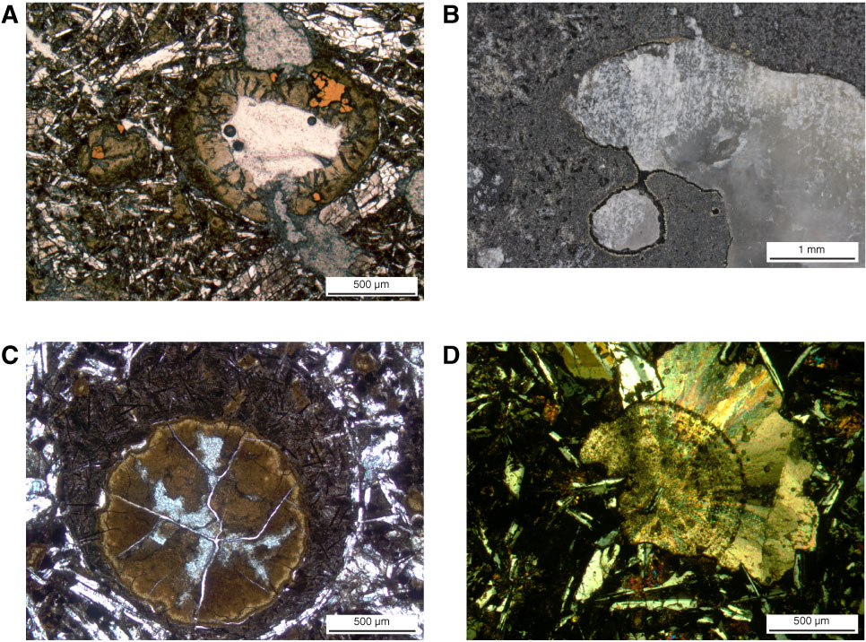
Figure F41. A. Photomicrograph of grayish green clay and golden brown pyrite partially filling a vesicle, Hole U1347A (Thin Section 79; Sample 324-U1347A-14R-1, 108–110 cm). Combined plane-polarized and reflected light. B. Close-up photograph of a vesicle lined with dark green clay and pyrite and filled with calcite (Sample 324-U1347A-12R-1, 30–31 cm). C. Segregation melt altered to dark brown clays and opaques around a brown clay vesicle (Thin Section 127; Sample 324-U1347A-23R-6, 95–96 cm). Plane-polarized light. D. Calcite with radial growth patterns in a vesicle (Thin Section 95; Sample 324-U1347A-17R-3, 65–67 cm). Cross-polarized light.
Previous | Close | Next | Top of page