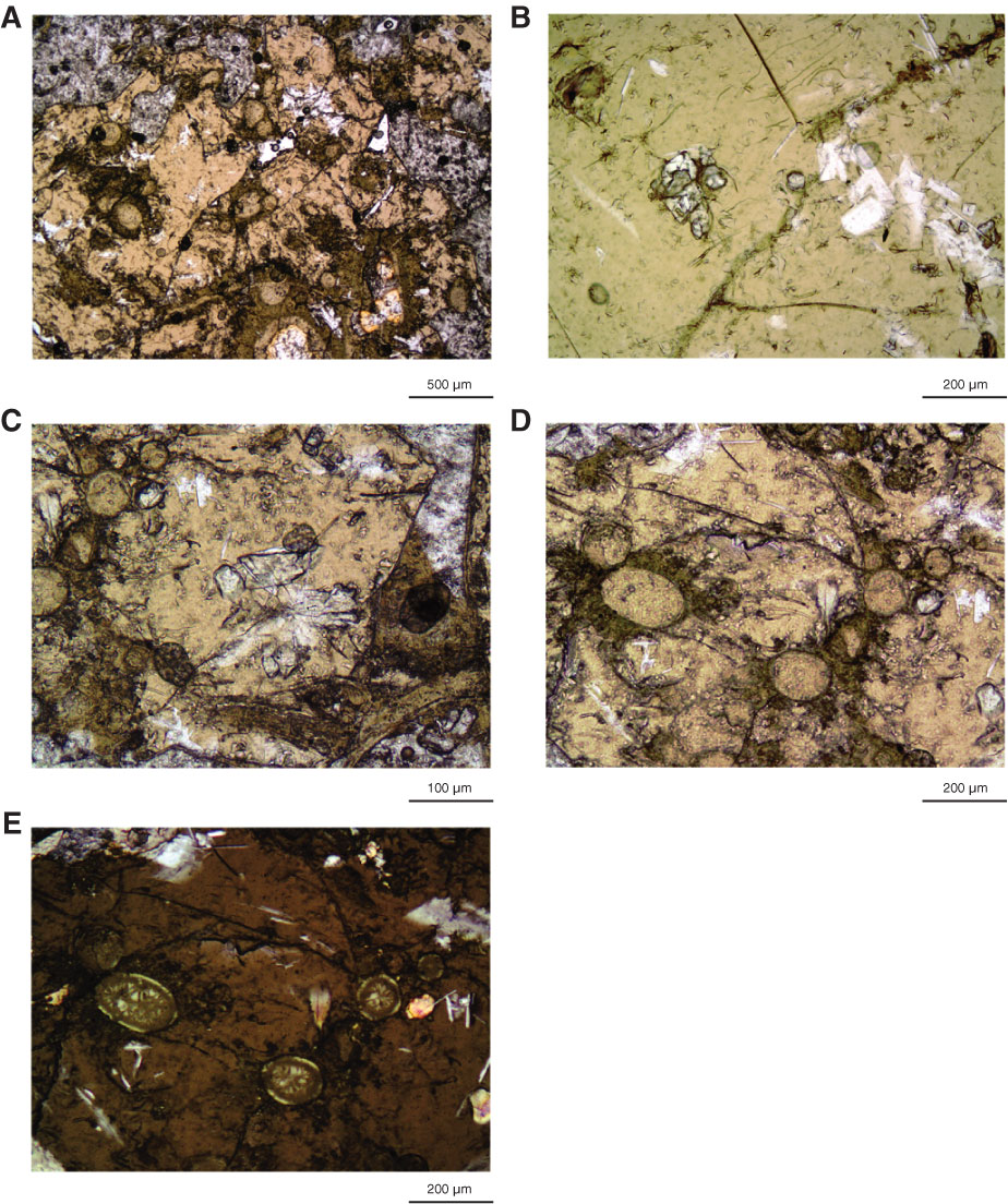
Figure F34. Thin section photomicrographs emphasizing details of spheroid formation in glass (Thin Section 197; Sample 324-U1348A-23R-1, 116–122 cm), Hole U1348A. A. Light brown areas of fresh glass. Altered areas contain fibrous orange clays along fractures and surrounding incipient spheroids. B. Fresh olivine (high relief) and intergrown plagioclase (white and elongate) and clinopyroxene (white and almost square) microlites. C. Larger bundle of intergrown plagioclase and clinopyroxene. D. Spheroids with almost the same color as the glass but surrounded by fibrous orange clays arranged along small cracks in the glass. E. Same view as D but emphasizing the radial pattern of fibropalagonite developing in the spheroids. Fresh plagioclase needles are white. A–D are under plane-polarized transmitted light; E is under partial cross-polarized light.
Previous | Close | Next | Top of page