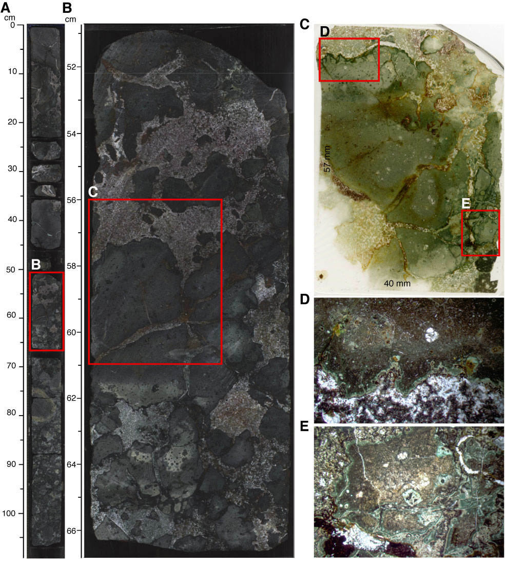
Figure F17. Core section images of (A) Section 324-U1349A-16R-6 and (B) interval 324-U1349A-16R-6, 56–61 cm; (C) thin section scan; and (D, E) photomicrographs of flow breccia in Unit V, Hole U1349A. Thin section scan location of C is indicated by the red box in B. Red boxes in C indicate photomicrograph locations. Width of field of view for D is ~15 mm (1.25x). Width of field of view for E is ~10 mm. Plane-polarized light.
Previous | Close | Next | Top of page