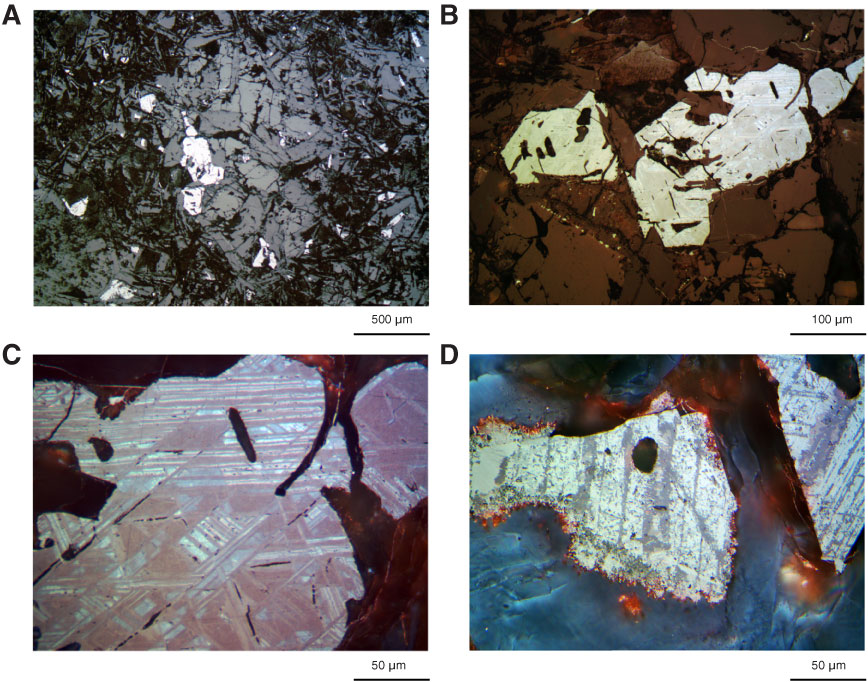
Figure F26. A–D. Photomicrographs of oxy-exsolution in coarse titanomagnetite at the rims of plagioclase-clinopyroxene ophimottles, Hole U1349A (Thin Section 219; Sample 324-U1349A-10R-2, 10–12 cm). In A and B, the titanomagnetite has skeletal morphologies with euhedral outlines. B and C show the same oxide mineral. The exsolution can be made out in air using lower magnification, as in B, but stands out in reflected light. Four different oxide minerals are present in both C and D. Exsolution lamellae are sharper and minerals have higher contrast in D, which is also more altered. Lamellae are raveled at the edges and evidently transformed to iron oxyhydroxides or hematite, which is very fine grained, not polished, embedded in the adjacent clinopyroxene, and only evident as internal reflections. Reflected light. C and D are with oil immersion.
Previous | Close | Next | Top of page