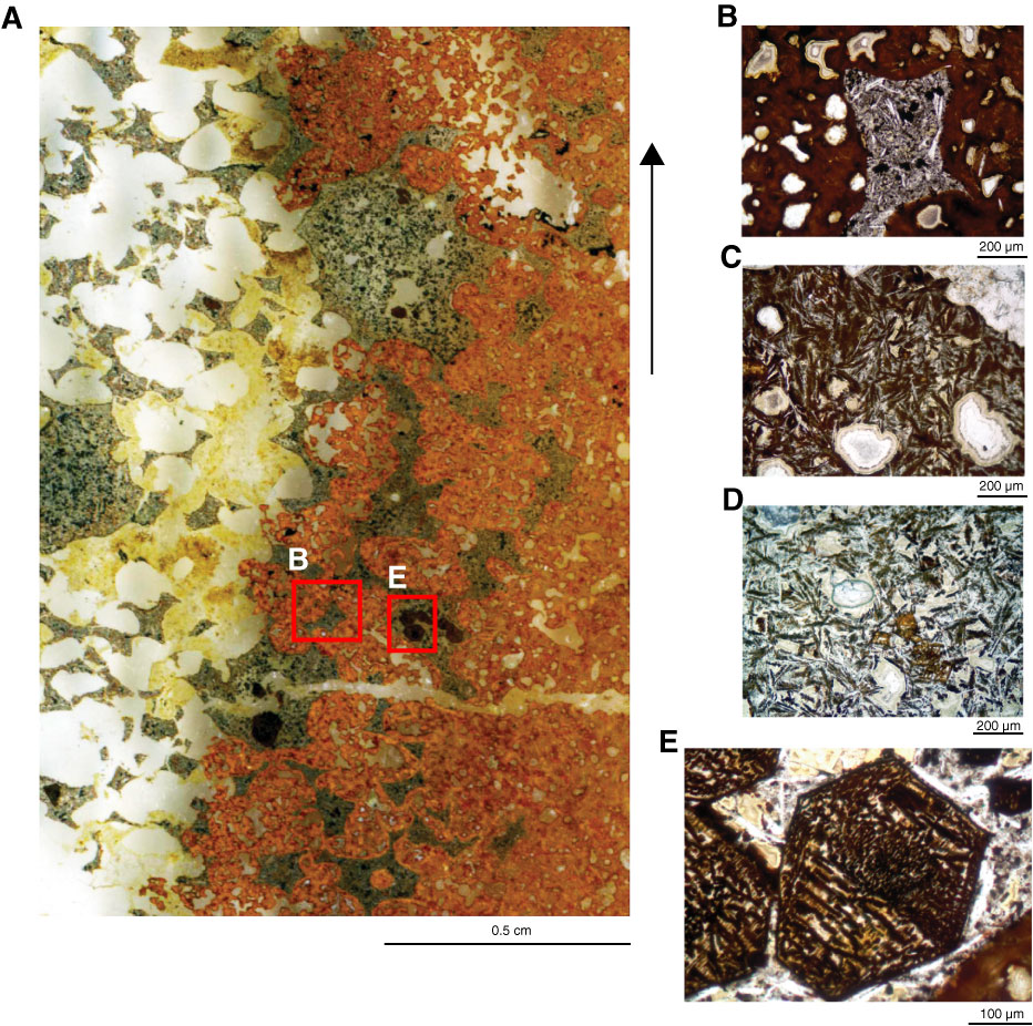
Figure F31. A. Partial thin section image, Hole U1349A (Thin Section 230; Sample 324-U1349A-11R-5, 11–14 cm). The two sides of the scan, orange and gray with white vesicles, are two different basalt compositions that cooled side by side and partially blended with each other in the variegated center of the scan. The material on the left is far more vesicular than that on the right, and in the central band, the orange-colored basalt filled vesicles of the gray. The gray material has ~75% vesicles, now filled with calcite. Arrow = orientation in core. B–D. Photomicrographs of the orange material in A at successively greater distances from blending zone, showing an increase of crystallinity of both plagioclase (white) and dendritic clinopyroxene (feathery brown, now altered). Photomicrograph location of B is indicated by red box in A. Transmitted light. E. Higher magnification image of one of the olivine crystals at lower center in A (location is indicated by red box). Crystal contains a trellised grid of hematite crystals, which are inferred to replace originally exsolved low-Ti magnetite during high-temperature alteration of the olivine.
Previous | Close | Next | Top of page