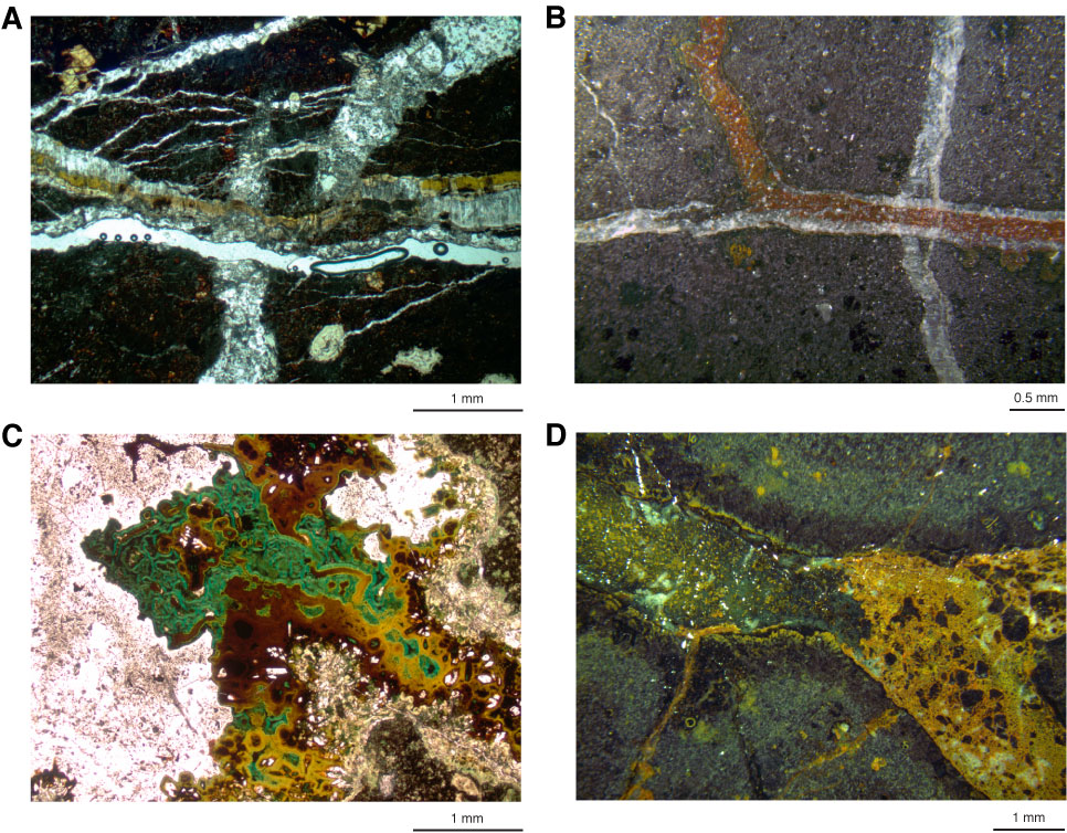
Figure F48. A. Photomicrograph of cross-fiber calcite vein containing saponite and cutting crystalline calcite veins, Hole U1349A (Thin Section 221; Sample 324-U1349A-10R-3, 43–48 cm). B. Close-up image of archive-half core with a calcite vein being cut by a vein of orange clays (Sample 324-U1349-14R-6, 48–49 cm). C. Photomicrograph of a vein in which Fe oxyhydroxides are overgrown by brown saponite, which is in turn overgrown by green nontronite and blue-green celadonite and subsequently is cemented by calcite (Thin Section 246; Sample 324-U1349A-14R-4, 50–56 cm). D. Close-up image of archive-half core with a vein in which green clays are being replaced by orange oxidized clays (Sample 324-U1349A-16R-5, 115–116 cm). A and C are under plane-polarized light.

Previous | Close | Next | Top of page