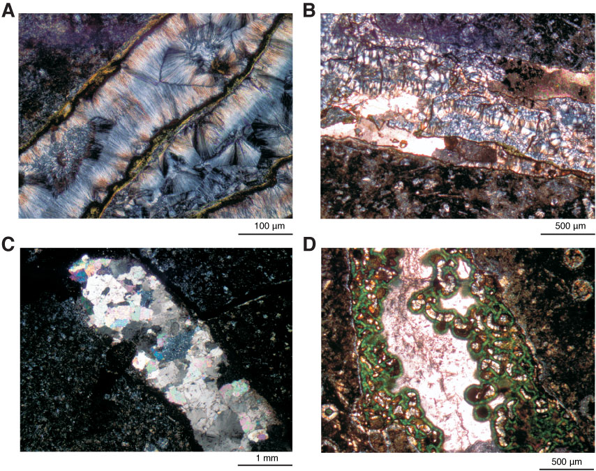
Figure F53. Photomicrographs of vein textures in Hole U1349A basalts. A, B. Cross-fibers composed of (A) needlelike and (B) spherulitic zeolites growing perpendicular to vein walls (Thin Section 260; Sample 324-U1349A-16R-6, 7–12 cm). C. Polycrystalline texture mainly displayed by calcite in veins (Thin Section 254; Sample 324-U1349A-15R-4, 104–110 cm). D. Intravenous vein texture displayed by disorderedly aligned calcites or zeolites (Thin Section 249; Sample 324-U1349A-14R-5, 53–54 cm). Cross-polarized light.

Previous | Close | Next | Top of page