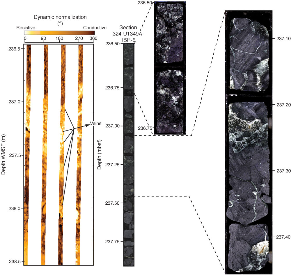
Figure F71. Formation MicroScanner (FMS) images of fractures and potential veins in the lower section of Hole U1349A. Core images provided to show similar structures in the core. Color levels on the core images were adjusted in order to accentuate the textures and contacts.

Previous | Close | Top of page