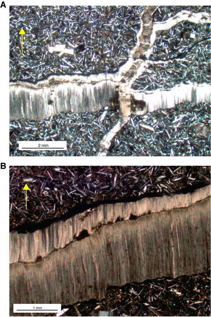
Figure F38. A, B. Photomicrographs of cross-fibers of needlelike calcites growing perpendicular to vein walls at (A) 1.25x and (B) 2.5x magnification, Hole U1350A (Thin Section 289; Sample 324-U1350A-16R-3, 71–74 cm). Yellow arrows = orientation in core. Cross-polarized light.
Previous | Close | Next | Top of page