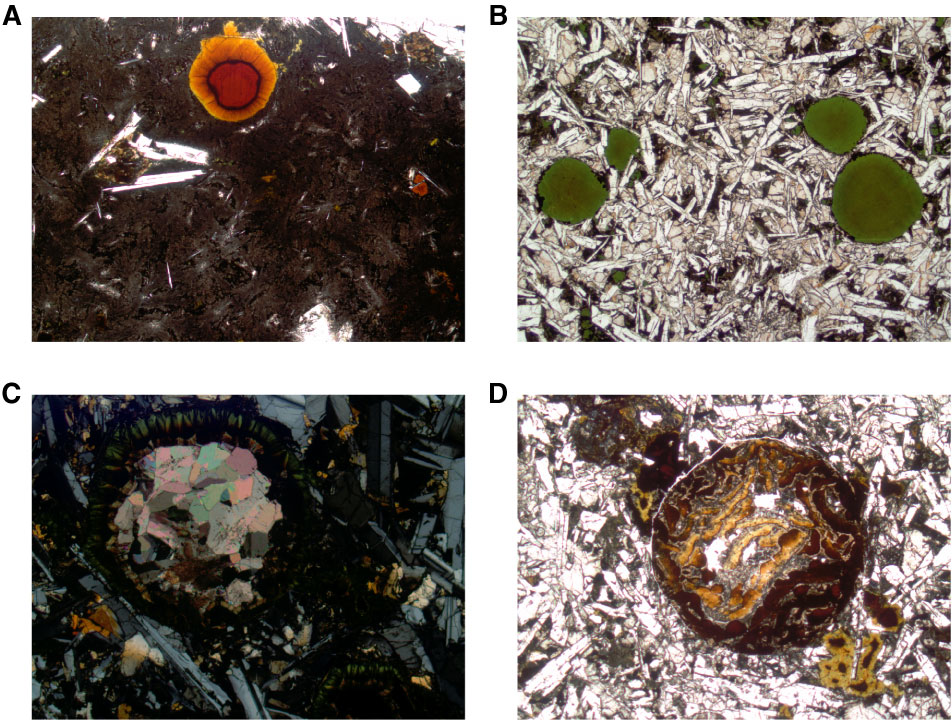
Figure F15. Photomicrographs of filled vesicles. A. Multilayered vesicle lined with bright orange iron oxyhydroxide followed by a saponite and iron oxyhydroxide mixed band and a deep red iron oxyhydroxide core (Sample 327-U1362A-7R-1 [Piece 5, 29–31 cm] (FOV = 2.5 mm; plane-polarized light). B. Vesicles filled with a mixture of saponite and celadonite with celadonite-rich rims (Sample 327-U1362A-9R-1 [Piece 20, 125–128 cm] (FOV = 4.5 mm; plane-polarized light). C. Vesicle with fibrous calcite lining and crystalline calcite core (Sample 327-U1362B-16R-3 [Piece 5, 89–91 cm]) (FOV = 2.5 mm; cross-polarized light). D. Multilayered iron oxyhydroxide and saponite-filled vesicle. Later alteration by saponite overprints initial layering to create concentric blebs of material. (Sample 327-U1367A-16R-1 [Piece 5, 89–91 cm]) (FOV = 2.5 mm; plane-polarized light).
Previous | Close | Next | Top of page