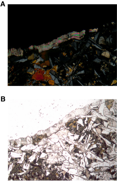
Figure F19. Photomicrographs of anhydrite present as a fibrous vein along edge of thin section (Sample 327-U1362A-18R-2 [Piece 3, 100–102 cm]). A. Cross-polarized light (FOV = 2.5 mm). B. Plane-polarized light (FOV = 2.5 mm).
Previous | Close | Next | Top of page