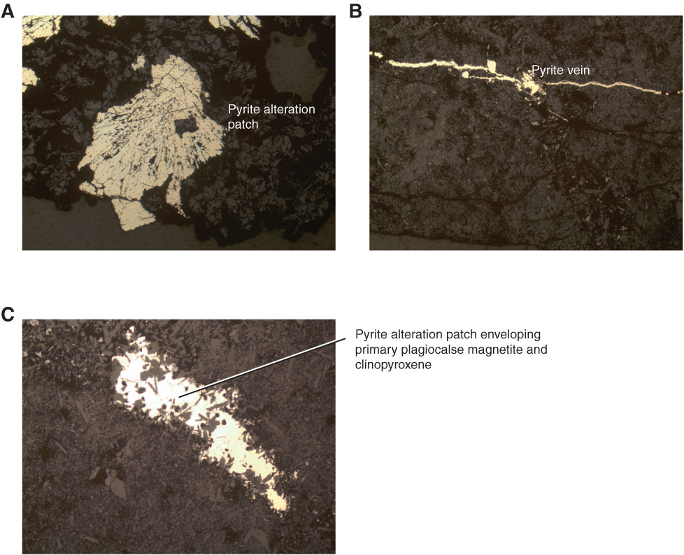
Figure F23. Photomicrographs of pyrite alteration. A. Pyrite alteration patch within a saponite and calcite alteration zone (Sample 329-U1365E-12R-4, 25–29 cm). B, C. Pyrite vein in B and pyrite alteration patch within a coarser grained portion of the groundmass in A (Sample 329-U1365E-12R-4, 70–72 cm). Reflected light. A and B are at 5× magnification and C is at 10× magnification.

Previous | Close | Next | Top of page