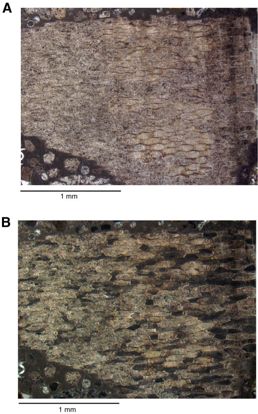
Figure F17. Thin section photomicrographs of inoceramid bivalve shell fragment showing honeycomb texture of prismatic shell layer (Sample 330-U1372A-5R-1W, 32–34 cm; Thin Section 8). A. Plane-polarized light. B. Crossed polars.
Previous | Close | Next | Top of page