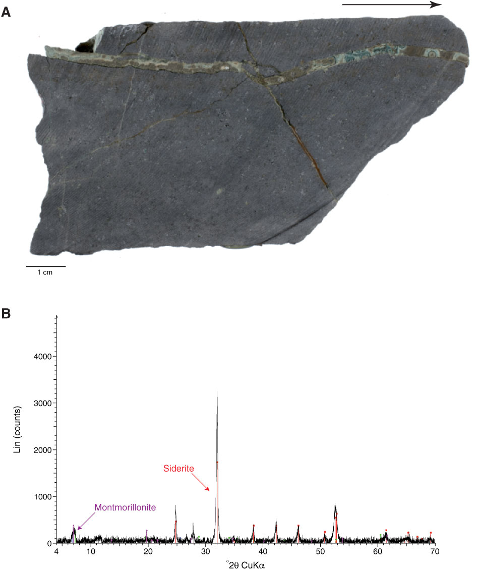
Figure F41. A. Close-up photograph of a composite vein consisting of siderite and brown and green clay (botryoidal aspect; nontronite, montmorillonite, and saponite) (Sample 330-U1372A-26R-1, 117–128 cm; dry surface; arrow points to top). B. X-ray diffraction spectrum for vein sample shown in A. The secondary mineral assemblage is predominantly composed of siderite with moderate montmorillonite. Minor peaks for nontronite and saponite are also present (not labeled at this scale).

Previous | Close | Next | Top of page