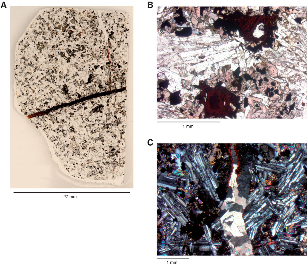
Figure F8. A. Close-up photograph of Thin Section 228 (Sample 330-U1375B-1R-1, 57–59 cm), showing a reddish-brown vein (from left to right) filled with goethite and another vein (from top to bottom) mostly filled with carbonate and some goethite (right-hand side of image). B. Thin section photomicrograph of completely altered microcrystals of olivine to hematite, iddingsite, and some Fe oxyhydroxides (moderately olivine-augite-phyric microgabbro) (Sample 330-U1375B-1R-1, 1–3 cm; Thin Section 227; plane-polarized light). C. Millimeter-size vein filled with carbonates and goethite on wall (upper part of image) (moderately olivine-augite-phyric microgabbro) (Sample 330-U1375B-1R-1, 57–59 cm; Thin Section 228; crossed polars).

Previous | Close | Next | Top of page