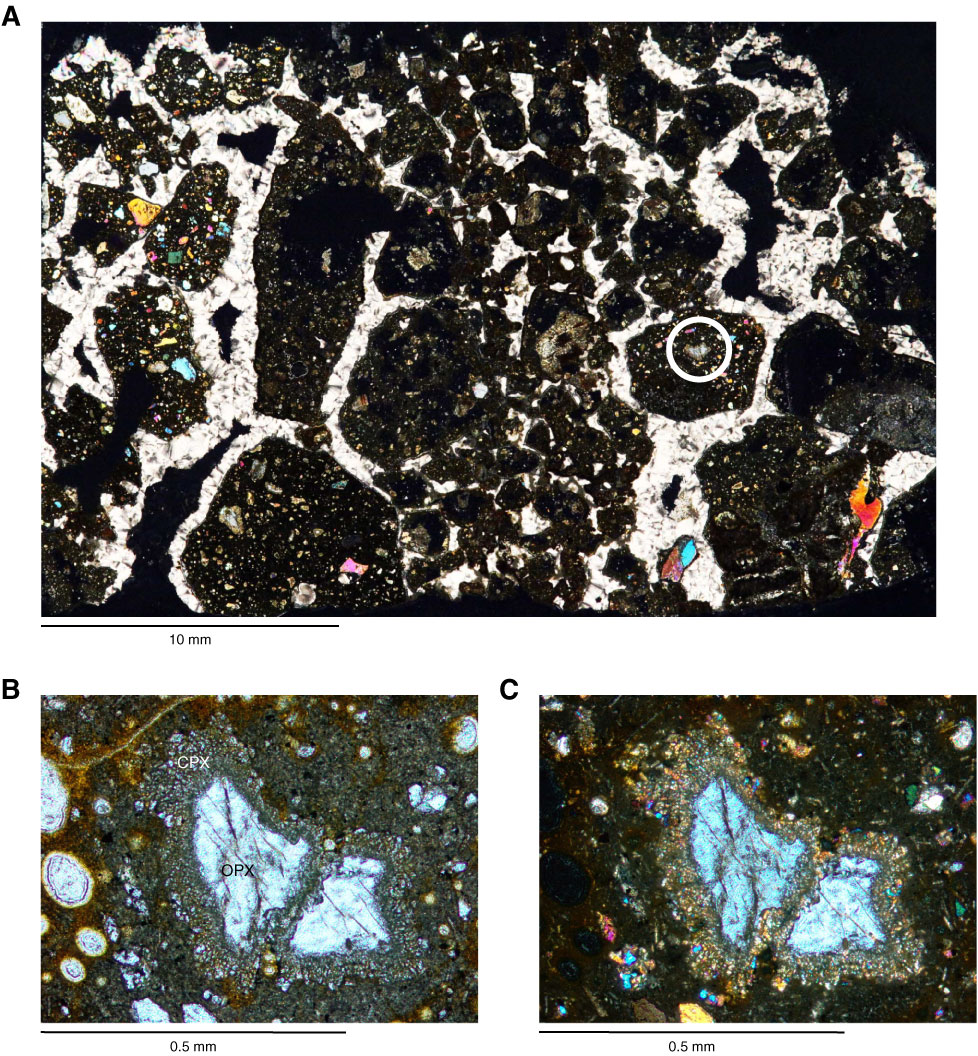
Figure F12. Thin section photomicrographs from Subunit IC (Sample 330-U1376A-1R-4W, 39–42 cm; Thin Section 234): (A, C) crossed polars, (B) plane-polarized light. Circle in A shows the location of B and C, which show an orthopyroxene xenocryst (OPX) surrounded by a reaction corona composed of tiny crystals of clinopyroxene (CPX). A scanned image of the archive half of this interval is shown in Figure F11.
Previous | Close | Next | Top of page