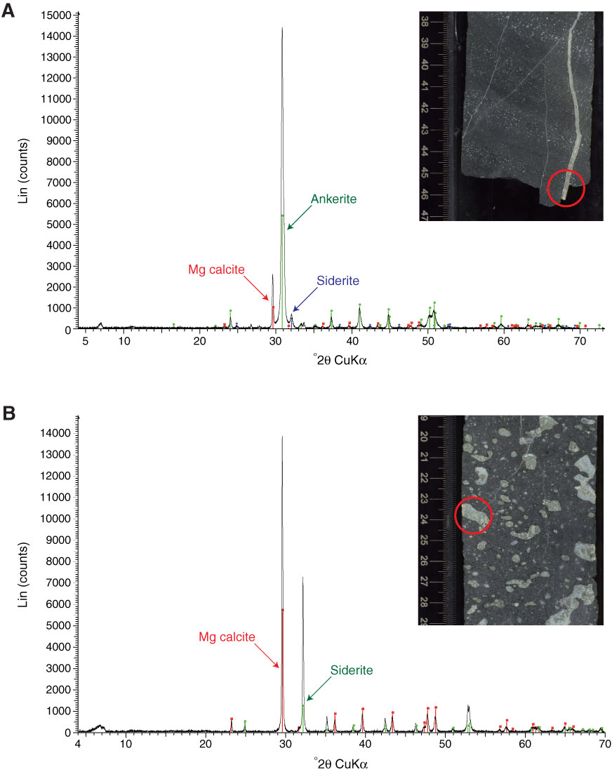
Figure F15. X-ray diffraction spectra and associated core photographs (analyzed zones are highlighted with red circle). A. Millimeter-thick vein filled with ankerite, Mg calcite, and siderite (Sample 330-U1377A-5R-2, 45–47 cm). B. Vesicle filled with Mg calcite and siderite (Sample 330-U1377A-6R2, 25–27 cm).
Previous | Close | Next | Top of page