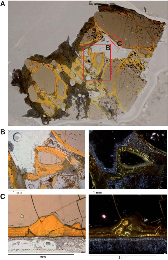
Figure F20. Photomicrographs of hyaloclastite, Unit 3 (Sample 336-U1383C-20R-1, 39–42 cm [Piece 28], Thin Section 50). A. Whole thin section photomicrograph showing partly palagonitized glass clasts with minor devitrification spherulites and microliths. B. Large red box in A: Concentric alteration rim of palagonite (yellowish brown) extending into fresh glass (light brown). A zeolite cement forms a thin (<100 µm) coating along palagonitized glass clasts, whereas the matrix is composed of a mixed assemblage of clay, palagonite, and zeolite. Left image is in single-polarized light, right image is under cross-polarized light. Cross-polarized image shows concentric palagonite bands with bright birefringence. Field of view (FOV) = 4 mm. C. Small red box in A: sharp transition between palagonite and fresh glass, which contrasts with chilled margin glass alteration (Fig. F18). The alteration front in this hyaloclastite remarkably lacks composite alteration halos and microtubules extending into the fresh glass. Note the thin external rim of zeolite at the bottom of the photograph with acicular to fibrous crystallization. Left image is under single-polarized light; right image is under cross-polarized light. FOV = 1.1 mm.
Previous | Close | Next | Top of page