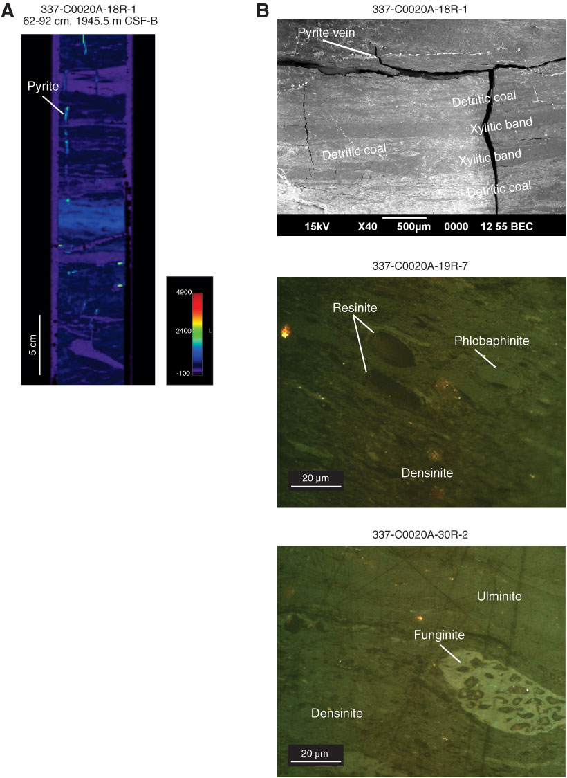
Figure F6. A. X-ray CT scan images from coal of Section 337-C0020A-18R-1. B. Top: SEM picture from a banded coal (xylitic and detritic bands) of Section 18R-1 with some pyrite veins (upper part). Middle: photomicrograph of coal from Section 19R-7; densinite with some resinite and phlobaphinite. Bottom: photomicrograph of coal from Section 30R-2; densinite with a large funginite in the lower part and ulminite in the upper third of the photomicrograph.
Previous | Close | Next | Top of page