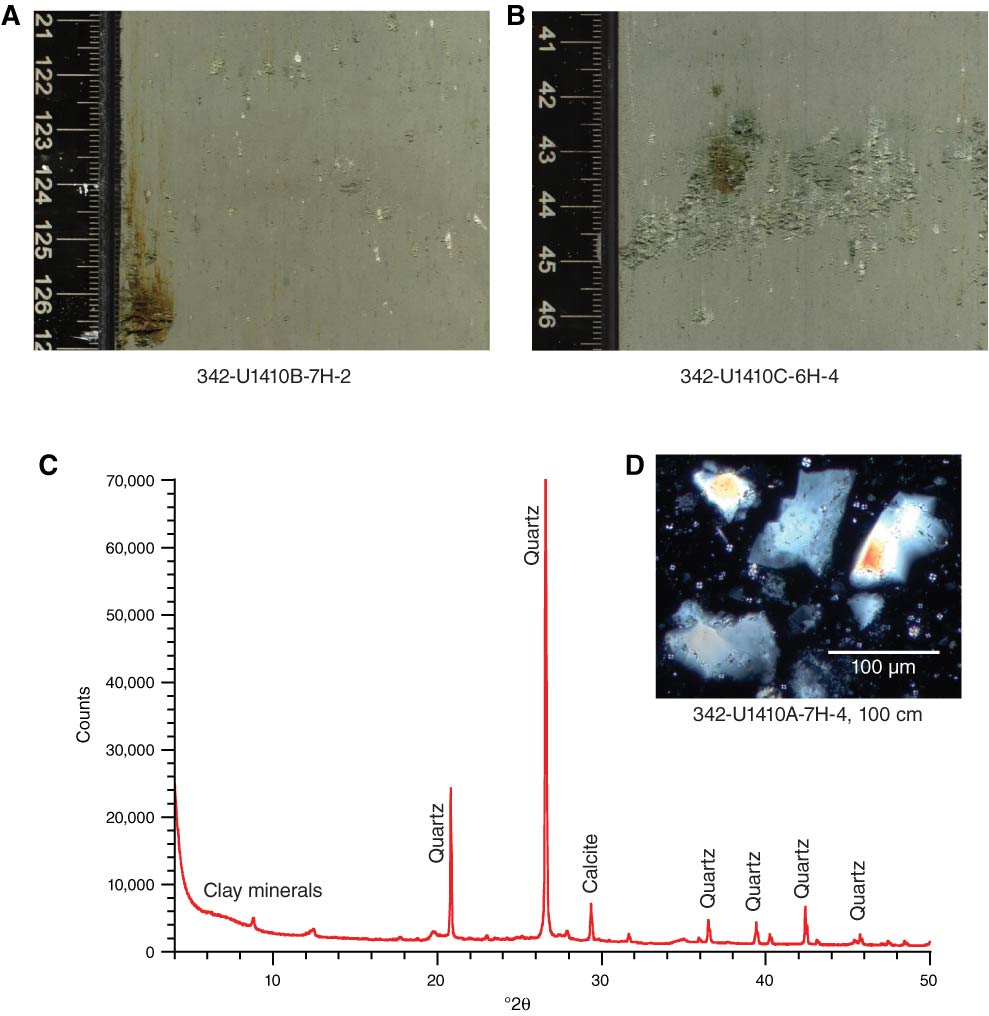
Figure F10. Core images of Miocene–Oligocene greenish gray nannofossil clay of Unit II showing dispersed quartz in (A) millimeter-scale patches/blebs and, more rarely, (B) a discontinuous lens, Site U1410. C. Mineralogy based on X-ray diffractometry of a sample of silt- to fine sand–sized white material. The clay and calcite peaks are from the background lithology within which the quartz occurs. D. Photomicrograph of smear slide showing angular quartz in coarse silt to very fine sand size range.

Previous | Close | Next | Top of page