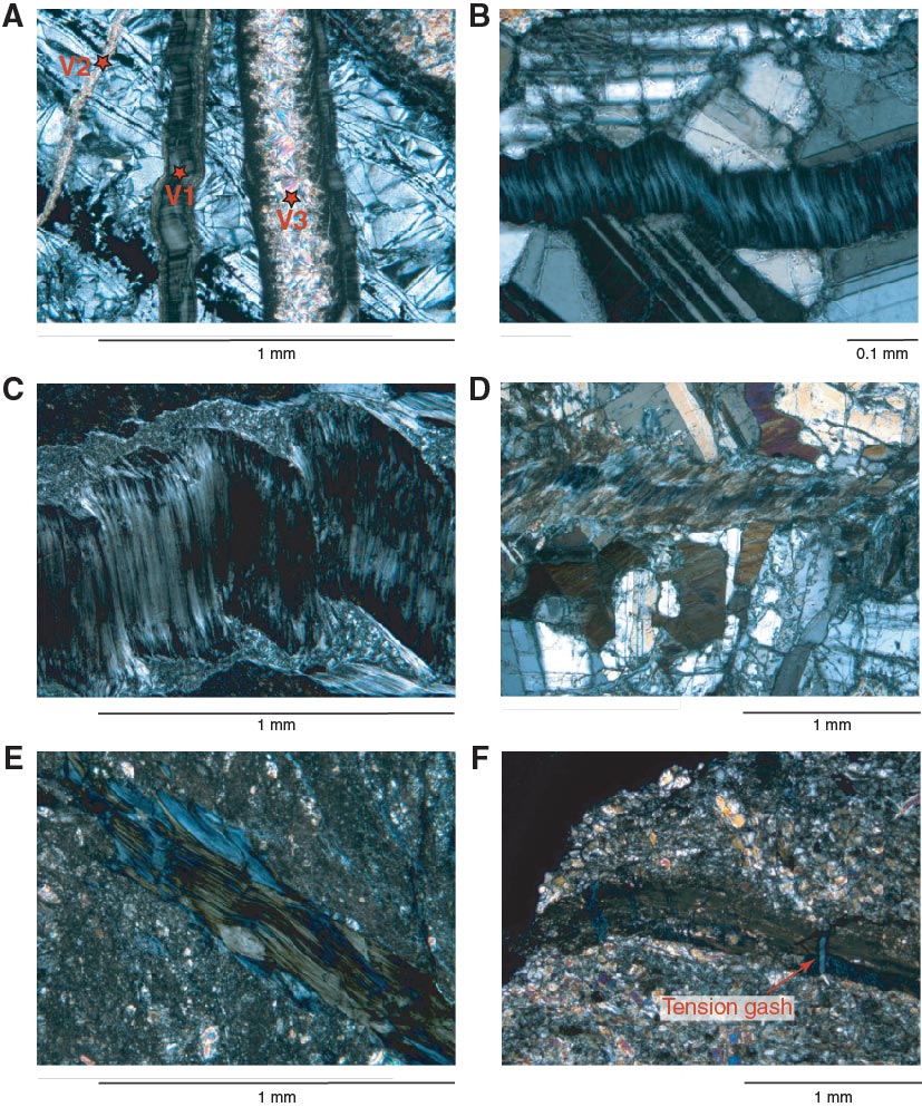
Figure F81. Microscopic character of veins (under crossed polars). A. Set of undeformed veins injected into serpentine (Sample 345-U1415J-19R-1, 18–19 cm [Piece 9]). V1 is filled with chlorite. Fibers grew normal to the vein wall (open crack). V2 is filled with prehnite, and V3 is filled with both chlorite and serpentine. B. Chlorite vein injected into weakly altered gabbro (Sample 345-U1415J-5R-1, 138–141 cm [Piece 18]). Fibers are perpendicular to the vein walls, but their slightly sigmoidal shape shows moderate shearing during or after crack opening and chlorite crystallization. C. Chlorite vein injected into altered troctolite (Sample 345-U1415J-18R-1, 141–143 cm [Piece 17B]). Fibers initially formed perpendicular to vein walls and were eventually sheared along the vein walls. Shear movement caused bending and displacement of the first filling material together with grain size reduction. D. Chlorite vein injected into moderately altered troctolite (Sample 345-U1415J-21R-1, 58–59 cm [Piece 9]). Fibers oblique to the vein walls and sigmoidal shape show that the crack opened and chlorite crystallized during shearing. E. Strongly sheared chlorite vein associated with cataclasite (Sample 345-U1415J-21R-1, 17–19 cm [Piece 4]). F. Chlorite-filled tension gashes filled with chlorite perpendicular to cataclastic foliation (Sample 345-U1415J-23R-1, 10–12 cm [Piece 3]).

Previous | Close | Next | Top of page