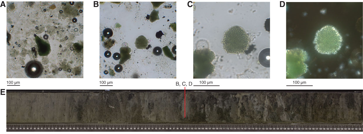
Figure F11. Smear slide images showing glauconite in (A) Sample 346-U1430A-6H-CC, 2 cm, and (B–D) in tephra layer at Sample 346-U1430A-9H-4, 100 cm. D is under crossed nicols and shows that glauconite has filled the interior space of a diatom but kept the diatom’s original texture. E. Photograph of full Section 346-U1430A-6H-CC.
Previous | Close | Next | Top of page