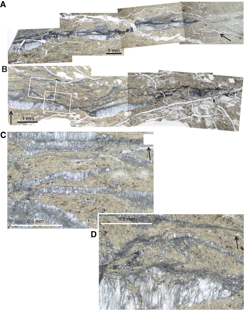
Figure F13. Photomicrographs of a vein formed of swarms of veinlets. A. Thin section TS 64–71 (open nicols). B. Thin section TS 78–85 (open nicols). C, D. Details of B (rectangles). Arrow = direction of core top.

Previous | Close | Next | Top of page