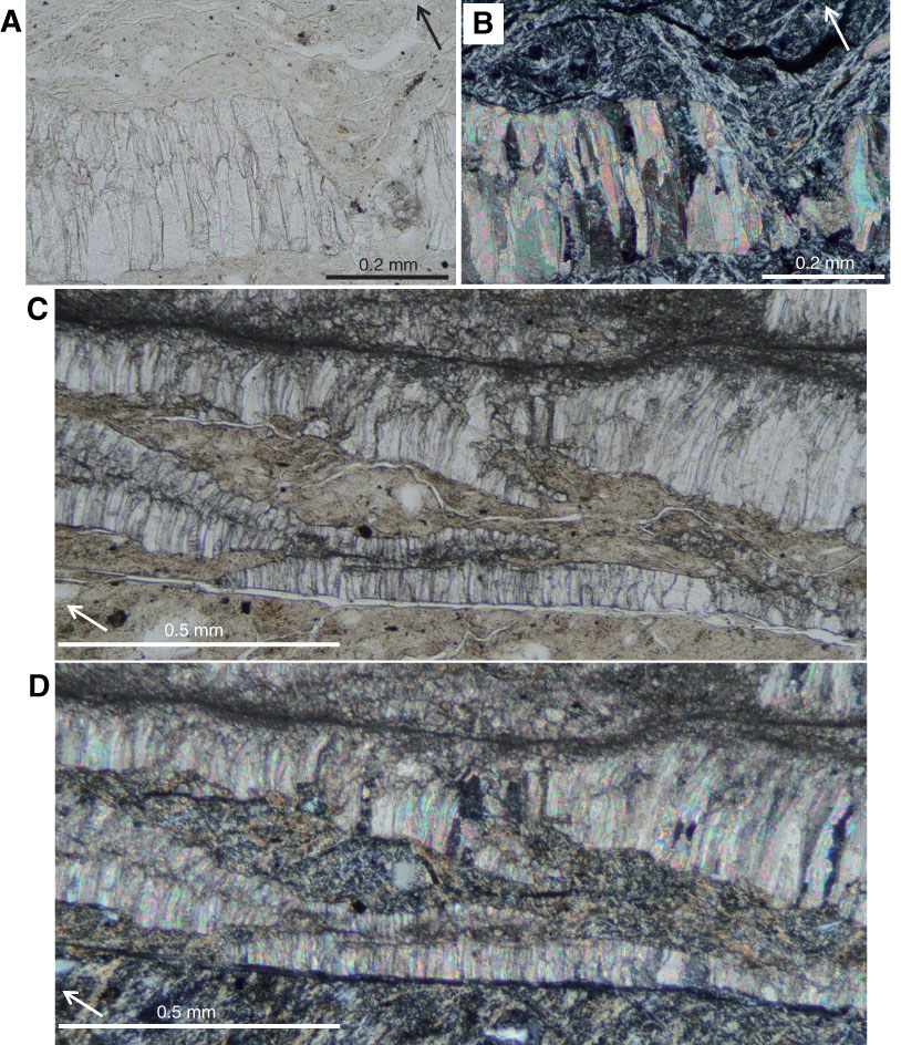
Figure F16. A, B. Photomicrographs of a microfault cutting a veinlet (thin section TS 71–78, open and crossed nicols, respectively). C, D. Photographs of wall rock inclusion bands and elongated calcite grains with broadening of grains toward the right (thin section TS 64–71, open and crossed nicols, respectively). Arrow = direction of core top.
Previous | Close | Next | Top of page