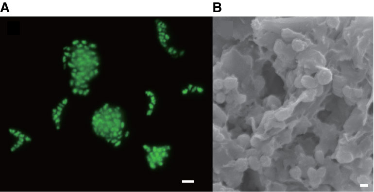
Figure F8. Microbial cells detected from methane hydrate–bearing sediments at 346 mbsf, Site C9001. A. Fluorescent microscopic image of SYBR Green I-stained cell aggregate. Scale bar = 2 µm. B. Scanning electron micrograph of cell aggregates. Scale bar = 200 nm.

Previous | Close | Next | Top of page