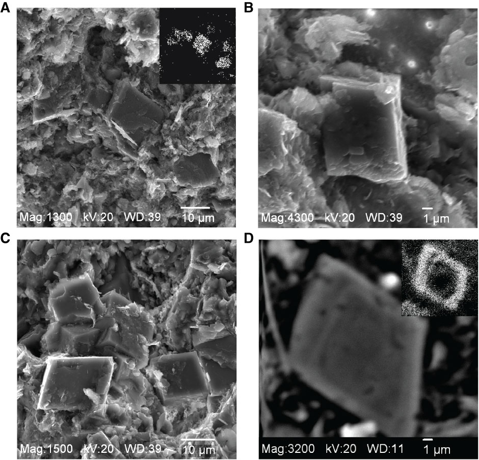
Figure F6. Electron micrographs of dolomite. A. Unconsolidated sediments (Core 307-U1317D-16H). Field of view shows relationship of dolomite rhombs with adjacent sediments; note abundance of clay mineral material. Upper right inset = electron dispersive spectrometer (EDS) “dot map” for Mg, confirming the location of dolomite crystals. B. Same as A, close-up of dolomite rhomb. Note growth features on surface of dolomite crystal and lack of identifiable structures of nannoplankton in micrite matrix suggestive of dissolution. C. Dolomite in a lithified sample (Core 307-U1318B-24H). Dolomite rhombs and associated sediment. D. Backscattered electron image of a sectioned dolomite crystal in a lithified interval of sediment. Note distinctive compositional zoning. Upper right inset = wavelength dispersive dot map of iron distribution.
Previous | Close | Next | Top of page