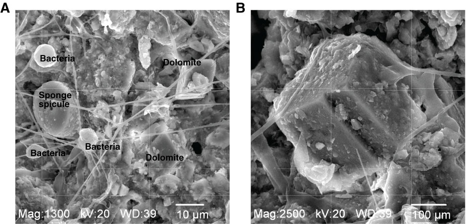
Figure F7. Electron micrographs of dolomite. A. Dolomite in an unlithified sample associated with microbial structures and filaments (Core 307-U1318B-24H). B. Close-up of upper right dolomite crystal in A with attached microbial filaments.
Previous | Close | Next | Top of page