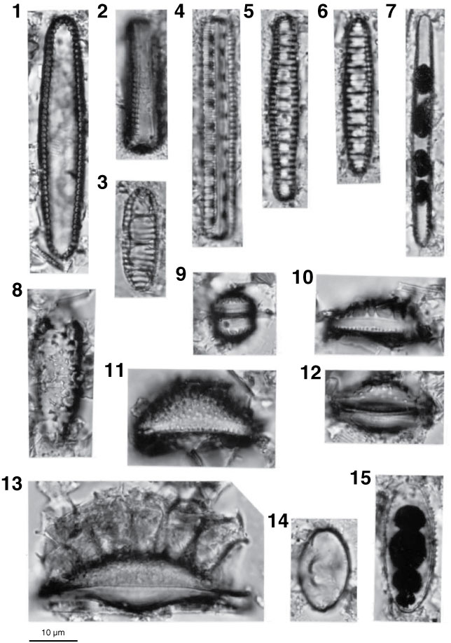
Plate P1. Light microscope images of marine diatoms, Holes U1325B, U1327C, U1328C, and U1329C. 1, 2. Thalassionema schraderi Akiba (Sample 311-U1329C-20X-CC), (1) valve view of valve and (2) girdle view of frustule. 3–6. Neodenticula seminae (Simonsen and Kanaya) Akiba and Yanagisawa; (3, 6) Sample 311-U1329C-5H-CC; (4, 5) Sample 311-U1325B-18X-CC; (4) girdle view of frustule. 7. Thalassionema nitzschioides (Grunow) H. and M. Peragallo (Sample 311-U329C-20X-1, 10–20 cm). 8–15. Chaetoceros resting spores and allied genera; (8–10, 12, 14–15) Sample 311-U1327C-20X-CC; (11, 13) Sample 311-U1328C-11X-2, 7–9 cm.
Close | Next | Top of page