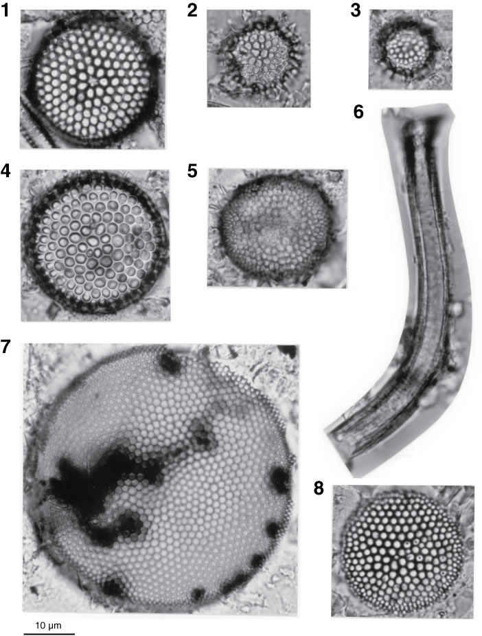
Plate P4. Light microscope images of marine diatoms, Holes U1325B, U1327C, and U1329C. 1. Thalassiosira antiqua (Grunow) Cleve-Euler (Sample 311-U1329C-20X-1, 10–20 cm). 2. Thalassiosira jouseae Akiba (Sample 311-U1329C-5H-CC). 3. T. jouseae Akiba (Sample 311-U1329C-16H-CC). 4. Stephanopyxis dimorpha Schrader (Sample 311-U1329C-16H-CC). 5. Actinocyclus oculatus Jousé (Sample 311-U1327C-32X-CC). 6. Proboscia curvirostris (Jousé) Jordan and Priddle (Sample 311-U1325B-18X-CC). 7. Actinocyclus curvatulus Janisch (Sample 311-U1329C-5H-CC). 8. Thalassiosira oestrupii (Ostenfeld) Proshkina (Sample 311-U1325B-18X-CC). Note many small grains of probable organic matter observed inside the diatom, which are also seen in other specimens (Pl. P1, figs. 7, 15).
Previous | Close | Top of page