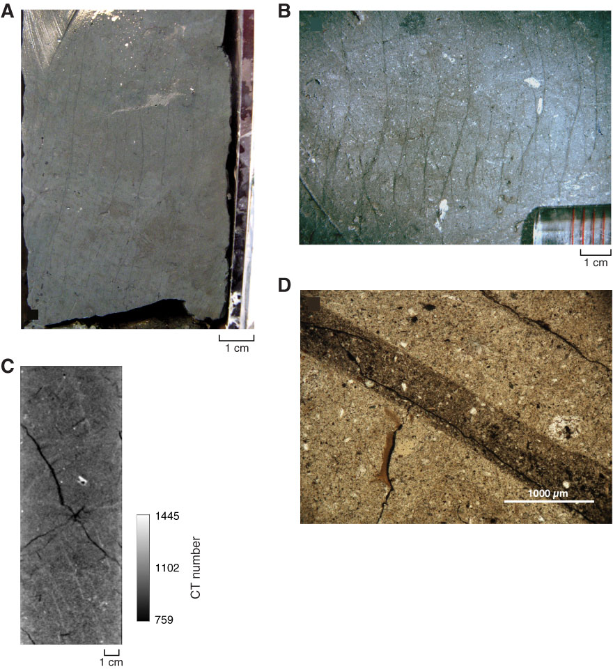
Figure F13. Vein structures. A. Archive core half (interval 315-C0001H-13R-2, 90–94 cm). Note black color and regular spacing between bands. B. Photomicrograph of slab section (interval 315-C0001H-13R-2, 90–94 cm). Veins are S-shaped, subparallel, branching, and anastomosing, sometimes with a normal offset of bioturbation features. C. CT scan image (Section 315-C0001F-15H-5). Veins are brighter, reflective, and therefore denser than the host rock. D. Photomicrograph of vein infill (interval 315-C0001H-11R-6, 94–102 cm) (plane-polarized light). Note darker color.
Previous | Close | Next | Top of page