Previous | Close | Next
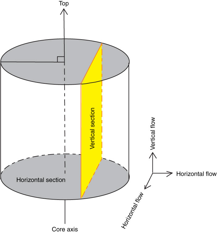
Figure F4. Diagram representing the horizontal and vertical section of core for imaging by environmental scanning electron microscope.
Previous | Close | Next | Top of page