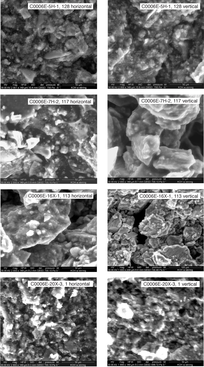
Figure F9. Environmental scanning electron microscope images for all specimens tested for permeability, Sites C0006 and C0007. Sections were cut parallel and perpendicular to core axis. See Figure F2 and Table T2 for bedding dips. (Continued on next two pages.)
Previous | Close | Next | Top of page