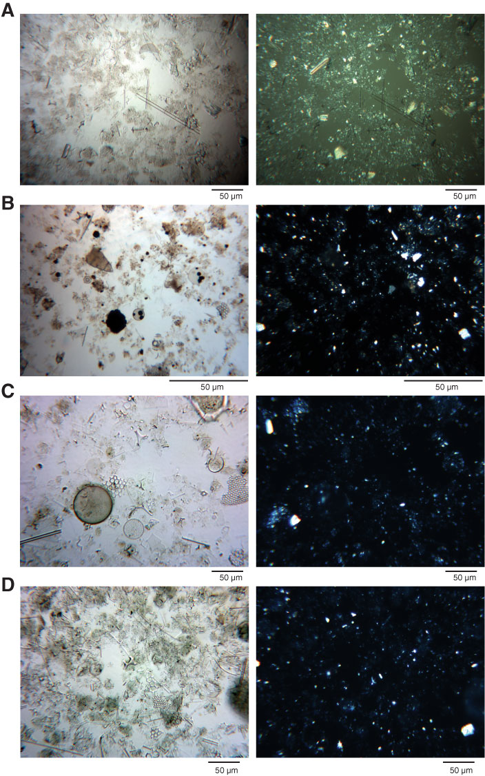
Figure F8. Light microscope photographs of smear slides indicating the dominant lithologies of Units I–III. Left image = plane-polarized light, right image = cross-polarized light. A. Nannofossil ooze with diatoms, Unit I (Sample 321-U1337A-4H-3, 95 cm). B. Diatom radiolarian ooze with nannofossils, Unit 1 (Sample 321-U1337A-5H-1, 98 cm). C. Diatom nannofossil ooze with radiolarians, Unit 1 (Sample 321-U1337A-5H-3, 127 cm). D. Radiolarian diatom ooze, Unit II (Sample 321-U1337A-12H-5, 131 cm). (Continued on next page.)

Previous | Close | Next | Top of page