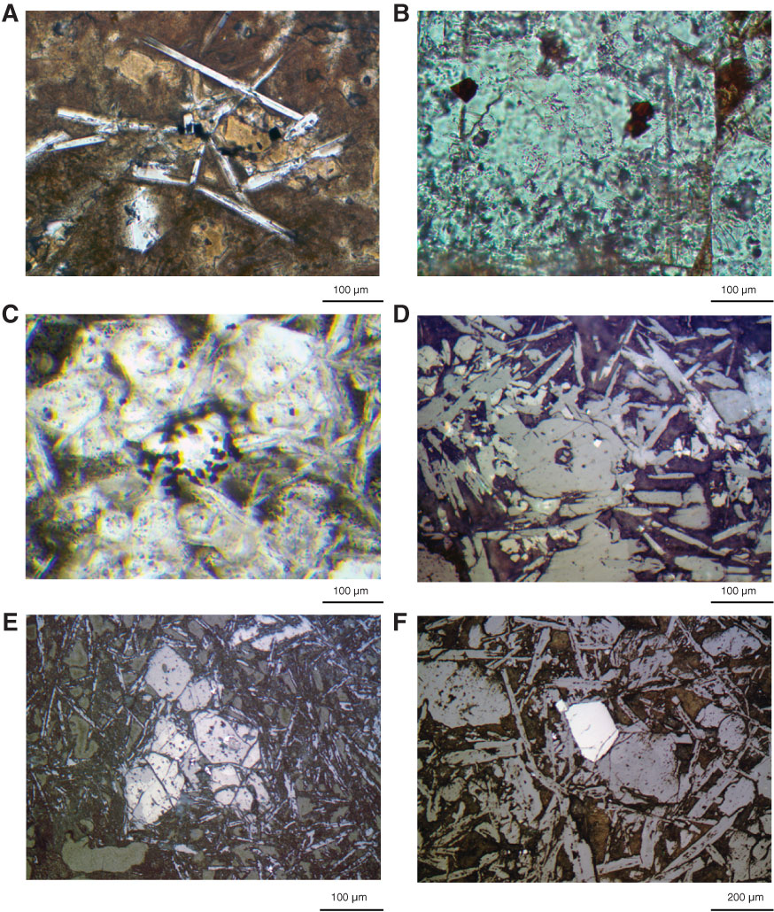
Figure F24. Photomicrographs of spinel. A. Small Cr spinel enclosed by or attached to olivine microphenocrysts, now pseudomorphed by clays (Thin Section 28; Sample 324-U1346A-8R-1, 56–60 cm). B. Small Cr spinel enclosed by or attached to olivine microphenocrysts, pseudomorphed by calcite. Engaging the conoscope to increase light reveals that spinel is dark brown (Thin Section 23; Sample 324-U1346A-7R-3, 67–70 cm). C. Clumped spinel (Thin Section 47; Sample 324-U1346A-13R-1, 102–105 cm). D. Spinel occurring within olivine (Thin Section 28; Sample 324-U1346A-8R-1, 28–29 cm). E. Thin Section 47 (Sample 324-U1346A-13R-1, 102–105 cm). F. Largest spinel in the core attached to an olivine pseudomorph on its right (Thin Section 36; Sample 324-U1346A-10R-1, 24–25 cm). A–C are under transmitted light; D–F are under reflected light.
Previous | Close | Next | Top of page