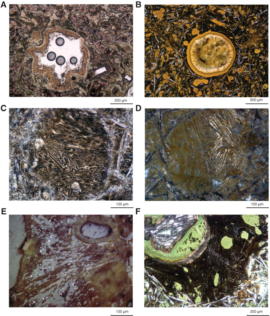
Figure F25. Photomicrographs of vesicles. A. Clay-lined vesicle in a near-glassy spherulitic zone but not a segregation vesicle (Thin Section 6; Sample 324-U1346A-4R-2, 23–26 cm). B. Segregation vesicle (Thin Section 53; 324-U1346A-14R-2, 107–108 cm). C, D. In C, the segregation vesicle is sliced laterally through a meniscus that did not intersect the vesicle. The slice shows an intergrowth of needles of clinopyroxene and tiny titanomagnetite crystals between the needles. D is the same sample, rotated and under cross-polarized light, showing that bundles of the clinopyroxene needles have common extinction angles and thus are dendritic intergrowths (Thin Section 50; Sample 324-U1346A-14R-3, 36–37 cm). E. Segregation vesicle with a portion of another meniscus and tiny skeletal titanomagnetite crystals randomly dispersed between clinopyroxene needles (Thin Section 37; Sample 324-U1346A-10R-2, 16–18 cm). F. Segregation vesicle showing a partial meniscus. The principal vesicle bubble at the upper left is lined with clays and calcite. The meniscus again consists of dendritic clinopyroxene and intergrown titanomagnetite, but in this case it is itself vesicular. As the main vesicle shrank during cooling, drawing melt into the original bubble, new bubbles in turn nucleated and grew within the meniscus. The newer vesicles are now filled with green clay (Thin Section 47; Sample 324-U1346A-13R-1, 102–105 cm). A–C and F are under transmitted light; D is under cross-polarized light; E is under reflected light.
Previous | Close | Next | Top of page